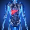
Description
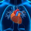
Cardiology
Our department is at the service of our patients every
day of the week and at any time of the day.
Reyap Hospital Istanbul has the opportunity to diagnose and treat all kinds of heart diseases
in all age ranges. Our hospital provides health care services to its patients
in the examination, diagnosis, treatment, rehabilitation, and coronary
intensive care in heart diseases.
All kinds of examinations, evaluations, and laboratory analyses required for diagnosis and
treatment for patients with heart disease are carried out in our outpatient
clinic, inpatient service, and coronary intensive care unit.
Cardiac Procedures
The cardiology department provides health care for adult
patients. Pediatric cardiology is a separate specialty.
Cardiologists are involved in the diagnosis and treatment
of patients with conditions such as;
Angina (chest pain caused by narrowing of the coronary arteries)
Arrhythmias
Heart murmurs due to valvular heart disease
Cardiomyopathy (heart muscle disease) with heart failure
including pulmonary
Edema
Coronary artery thrombosis and myocardial infarction
Arterial diseases (atherosclerosis, arthritis, atheroma)
Joint care of pregnant women with heart disease with their babies
Types of congenital heart disease, such as arterial/ ventricular septal defect
Cardiologists also must improve survival rates and
quality of life following a heart attack, heart failure, or arrhythmias.
Outpatient clinic examinations
Examinations are carried out by our physicians between
08:30 and 17:30, 6 days a week (except Sunday).
Electrocardiography (ECG/ EKG)
It is the recording of the electrical activities that occur in the heart to examine the functioning
of the heart muscle and the neural conduction system. ECG is one of the most
important diagnostic methods in the diagnosis of heart diseases. It has
essential importance especially in the diagnosis of cardiovascular occlusion,
rhythm disturbances, heart valve diseases, and heart failure.
Treadmill Stress (Exercise) Test
The treadmill stress test investigates the presence of
cardiovascular disease, determines the effectiveness of the treatment,
determines whether the arrhythmia occurs because of stress, examines the stress
capacity of the patient in various heart diseases, questions the effects of
stress on blood pressure in patients with hypertension and helps to determine
the operation time in valvular diseases.
During the stress test, the patient walks on a treadmill. The speed and inclination of the floor
gait are adjusted by the doctor to increase the heart rate. The exercise test
is an examination performed by the method of taking ECGs serially and measuring
the blood pressure while the person is walking on the treadmill at certain
speeds. Meanwhile, the patient’s complaints, heartbeats, and blood pressure are
monitored, and data are recorded by continuous ECG monitoring. It enables the
detection of abnormal findings that cannot be detected in ECG at rest, after
exercise. The stress test requires the attendance of medical staff from the
beginning to the end. Effort test provides great convenience in the early
diagnosis of heart diseases. The result is given immediately after the
examination.
Transthoracic Echocardiography
Echocardiography is the examination of heart structure
and performance by sound waves (ultrasound). The sound waves that are sent to
the heart and reflected from the heart are analyzed in the echocardiography
device, and information about the heart’s muscle tissue, heart valves, and
large vessels emerging from the heart is obtained.
Since it is not a method that involves radiation, and no medication is used during the procedure,
it can be applied to anyone, including pregnant women and newborn babies,
without any side effects and pain. During the procedure, the patient is asked
to sleep on a stretcher. After a water-based gel is applied and a probe, which
is the part that sends the sound, is placed doctor holds the device in various
positions in the chest area, allowing the heart to be examined. Very detailed
measurements and analyzes are made on these images.
Diagnosis of all structural diseases of the heart (heart enlargement, heart muscle diseases,
heart valve diseases, hypertensive heart diseases, heart failure, heart
membrane diseases, heart masses, clots, heart tumors, congenital heart
abnormalities, even the largest diameter vein in the body called aorta
diseases) is possible with echocardiography procedure. No preparation is
required for echocardiography. The result is given immediately after the
examination.
Stress Echocardiography
Stress echocardiography is used to investigate whether
there is an obstruction or constriction in the vessels feeding the heart
(coronary vessels), to decide whether a treatment other than drugs is required
in patients with a heart attack (myocardial infarction), and to understand the
severity of the condition in valvular heart diseases. It is a safe and
easy-to-apply technique that provides very important information.
Stress echocardiography is performed by simply examining the heart with sound waves before
and after the stress. The patient doesn’t feel pain during the procedure.
Stress is being created by the Treadmill device or by applying certain drugs to
increase the workload of the heart. The result is given immediately after the
examination.
Transesophageal Echocardiography (TEE) Test
The heart is located in the rib cage just above the
esophagus. TEE measurement is a method used when the echocardiographic
recording of sufficient quality cannot be made due to the chest structure of
the patient (lung disease, deformity, etc.) or when it is necessary to evaluate
the intra-cardiac formations more closely.
Transesophageal echocardiography is a procedure that enables the visualization of some regions
that cannot be visualized with conventional ECG and a more detailed
echocardiographic evaluation performed from the esophagus. A very clear,
detailed image is obtained with a thin tube (probe) that is lowered into the
esophagus through the mouth. It takes 30 minutes with preparation before the
procedure. The result is given immediately after the examination.
Rhythm (ECG) Holter
Rhythm holter is a device consisting of 3-4 cables and
electrodes which are placed on the chest. It provides long-term follow-up of
the heartbeat during daily life, and thus, the entire electrical activity of
the heart during the day is recorded. After a daily recording, the doctor takes
off the device from the chest and analyzes the records.
The biggest advantage of rhythm holter is that it can find the source of symptoms such as
palpitations and pain that do not occur during the examination but occur during
the day. The patient continues to do the routine activities during the day
while the holter device is working. Thus, heart rhythm disorders can be
analyzed and diagnosed, as well as how effective the treatment is. During the
recording, the patient is asked to repeat the events (drinking coffee,
exercise, etc.) that increase the complaints. Registration is usually 24 hours
but can take up to 48 to 72 hours as per the doctor’s request.
Another function of the rhythm holter is to evaluate the pacemaker function in patients with
pacemakers.
Event Recorder
The event recorders, which have the same operating
principle as the rhythm holter device, are used in the diagnosis of rhythm
disorders that develop less frequently. In addition to being worn on the chest
for 14 days, it can be placed under the skin for 6 months to 1 year. Recording
time can be controlled and the device can only record when symptoms occur.
Blood Pressure Holter
The blood pressure holter enables the diagnosis of
hypertension and the efficiency of its treatment by recording the blood
pressure and pulse of the person at regular intervals for 24 to 72 hours.
During the recording, the cuff of the sphygmomanometer is tied to the arm, and
blood pressure and heart rate are recorded during routine activities (activity,
sleep, rest, etc.) throughout the day. By analyzing this data on the computer,
the moments when blood pressure elevates and drops are found. Thus, activities
that increase symptoms are understood. In addition to providing early
diagnosis, blood pressure holter is also useful for planning a treatment plan.
Tilted Table Test
The tilted table test is a test that investigates the causes of fainting as a result of changes
in blood pressure and/or arrhythmia caused by sudden movements after prolonged
standing, standing still, or sitting. It is used in the differential diagnosis
of fainting. During the test, the patient lies on the table and the table moves
to an upright position. An excessive drop in blood pressure or arrhythmia in
heart rate indicates a cardiovascular disease.
Coronary Angiography (CAG)
Coronary angiography is a device that diagnoses narrowing
and obstruction in the arteries feeding the heart. For the test, arteries in
the groin or arm are used as intervention points. Thanks to the dye sent from
the intervention site, the overall view of the vein is obtained. Thus,
constriction, dilation, obstructions, deformities, and congenital or acquired
problems throughout the vessel are detected.
The patient’s hospitalization is required for the coronary angiography test. The patient
needs to rest for 4-6 hours after the procedure, and this period is longer for
patients who have been applied bypass before, patients who have undergone
various previous cardiac surgeries, and patients with occlusion in the inguinal
or arm vessels.
Percutaneous Transluminal Coronary Angioplasty
(PTCA) – Stent
PTCA (stent) is the method used in the treatment for
narrowing or complete occlusion in the vessels supplying the heart detected
after coronary angiography. Like coronary angiography, PTCA is performed in the
angiography laboratory without general anesthesia, using the same sheath that
was placed in angiography. The processing time is variable. At the end of the
procedure, the patient is taken to the proper service according to the doctor’s
guidance.
Coronary balloon angioplasty is performed using specially designed materials. First, the
catheter is placed through the sheath placed at the intervention site, and the
vessel is passed through the stenosis area with a very thin guidewire advanced
through this catheter. The balloon is slid over this guidewire and delivered to
the diseased location. Later, this balloon is inflated by giving liquid from
the outside, and therefore the stenosis disappears.
During this enlargement (swelling), the person may feel chest pain. This inflation and
lowering procedure may need to be repeated several times for some severe cases.
In the subsequent control, the process is terminated when it is determined that
the opening is sufficient. It is rare to ensure a smooth opening. Besides,
stents are preferred in 95% of patients to reduce the risk of restenosis in the
future.
The stent is mounted on the balloon in the form of a thin metal wire. The stent, which is tightly
attached to the balloon, enlarges when the balloon is inflated and expands to
the vessel wall, and remains enlarged there. Thus, the narrowing of the vein
due to its flexibility is prevented. In the past, only stents made of stainless
metal were used, but today, depending on the technological progress, new and
different stents with drug release and soluble quality are used. The doctor
decides the type of stent he is going to use by considering the patient’s needs
and wants.
Since large amounts of blood thinners are used during the procedure, the catheter in the groin or
arm is not removed immediately but is removed after waiting for a while.
PTCA is an application that requires one-day hospitalization. After one day the patient is
discharged if the doctor determines it’s appropriate. Patients need to stay
away from stressful environments and sexual intercourse for 15 days. Patients
need to be in their home for 2 days, and they should take 15 days of work to
rest. The doctor decides on the date when the patient travels by plane and goes
on a road trip.
While coronary angiography is routinely performed from an artery in the groin called the femoral
artery, in our clinic, this procedure can also be performed from an artery in
the wrist called the radial artery. Besides, in our clinic, occlusion of not
only heart vessels but also vessels such as kidney and leg vessels called
peripheral vessels can be treated with balloon and stent methods.
Cardiac catheterization
Cardiac catheterization is a method performed by groin and arm arteries similar to
coronary angiography procedure but often requires simultaneous vein
intervention. Cardiac catheterization is a diagnostic method used in the
diagnosis of congenital or subsequent diseases related to the structure of the
heart, anomalies, congenital or subsequent heart holes, and whether a different
treatment is required. The blood sample is taken from each heart cavity entered
and the pressures are measured. If necessary, a screening test is performed
from these gaps by giving dye. Thus, it is determined whether the heart
cavities and vessels are enlarged or whether there is a transition between the
cavities in the heart.
Cardiac catheterization is performed in patients who are believed to have a hole in the
heart and whose echocardiography is diagnosed, and necessary information for
the preoperative phase is determined. Defects in the functioning of the heart
valves and walls can also be demonstrated with a heart catheterization. The
cardiac catheterization procedure takes about 30 minutes. After the procedure,
the sheaths placed in the arteries and veins are pulled and the bleeding is
stopped with external pressure. The patient is taken to the appropriate service
by applying a tight bandage to that area. 6 hours of rest is usually sufficient
after the procedure. After 6 hours, if the doctor believes it appropriate, the
patient can be discharged. Severe complication risk is extremely low in cardiac
catheterization and coronary angiography.
Peripheral Angiography and Angioplasty
(Visualization of leg, neck, and arm vessels)
The technique of performing these procedures is similar
to imaging the heart vessels and enlarging the existing stenosis. However,
while other procedures treat the coronary arteries (the vessels that feed the
heart) peripheral angiography enlarges the stenosis in the larger vessels of
the body such as the arm, leg, or neck vessels. Since the veins are wider, the
materials used here are also in different sizes respectively.
Temporary Pacemaker Implantation Procedures
(Single or, Double Chamber Pacing)
In case the heartbeat slows down due to the insufficient
speed of the simulation center in the heart, or the inability of the stimulus
to be transmitted from the brainstem, the patient needs implantation of
pacemakers to regulate the heart rate to maintain a healthy life. The procedure
is usually performed under local anesthesia by placing thin wires called
electrodes through the large veins leading to the heart in the neck, chest or
groin, and connecting it to a generator outside the body. This procedure can be
done at the bedside or under an x-ray machine. The process usually takes 20-30
minutes. When the temporary battery requirement is removed, the wire placed
inside the heart is taken out.
Pacemakers
Millions of people around the world have implanted a pacemaker.
These high-tech small devices are used for many purposes, from preventing slow
heart rate to treating heart failure, acting as a pump for the heart, and
preventing sudden deaths. The device, which eliminates the complaints
experienced after insertion, helps the patient return to their regular life by
increasing the quality of life. There are 3 types of pacemakers:
One-wire and 2-wire batteries to prevent slow heart rate
3-wire batteries (KRT) used to treat heart failure
Defibrillators (ICD)
that give electroshock if the heart cannot perform its pump function due to the
arrhythmia
Pacemakers are
implanted in people who have heart arrhythmia and cannot manage their lives
normally. These patients can return to their regular life with the help of a pacemaker.
People with pacemakers can go back to work, do household chores, drive, travel,
swim, continue their hobbies, and sex lives.
People with pacemakers should carry their pacemaker identification cards with them at all
times. While traveling, they should learn the nearest clinics in their
destination. After the pacemaker is inserted, its performance must be
monitored. The pacemaker, which is a small computer, can be read from the
outside with the help of another computer using a method called the telemetric
method. In this way, information such as how the patient’s heart rate
progressed, how long the pacemaker worked, did he or she experienced arrhythmia
from time to time, was always connected to the pacemaker, or if there were
other rhythm disorders. Besides, it is possible to externally program the
pacemaker how many volts the battery should work or the values that should keep the heart
rate regular. The patient must go for a check every 6 months for 7 years, which
is the average battery life. These examinations are very important as it can be
predetermined that the battery will run out. All brands and models of batteries
can be monitored in our hospital.
Electrophysiological Studies (EPS)
It is an interventional diagnosis and treatment method
performed by placing thin cables called electrode catheters into the heart
through thin sheaths placed in the inguinal vessels in the
electrophysiology/angiography laboratory. The electrical signals received
directly from the heart are evaluated by advanced computers and deviations are
investigated. In this way, it can be understood whether the main center
stimulation system of the heart works properly and whether the system that
transmits the signals function well.
In patients with heart palpitations, often in the form of rapid beats, the rapid beats, which
are the cause of the patient’s complaint, are created with the stimuli given
from these cables placed in the heart with special methods and the reasons for
their incident are investigated. When short circuits are detected, tachycardia
can be completely treated by applying special current point energy consisting
of radio waves. In this way, permanent treatment of most of the fast heartbeat
palpitations has become possible today. Electrophysiological examinations performed
for diagnostic purposes take 30-60 minutes. If a therapeutic intervention is
required, this procedure can take up to 1 to 4 hours.
Catheter Ablation
It is the treatment of arrhythmia by giving radio waves.
This method is used in rhythm disorders that cannot be managed with drugs or
when patients do not want to take medication for life. In some cases, the
rhythm disturbance can be so severe that it can be life-threatening. In such
cases, a direct catheter ablation method may be required. The procedure is
performed by numbing the needle insertion sites with local anesthesia and, in
some cases, under general anesthesia.
During the procedure, sedative medication can be used to make the patient feel
comfortable. The success rate of the treatment of rhythm disorders in the form
of the fast beating of the heart with catheter ablation varies between 70-100%
depending on the type of palpitation targeted to treat and the location of the
short circuit. The success of this treatment is for patients to never experience
palpitation again. The probability of the flutter recurrence after successful
application varies according to the type of arrhythmia. For example, this
possibility is between 3-5% in palpitations due to short circuits in the heart.
In our hospital, in addition to the radiofrequency ablation method called the
“burning” method, the “freezing” method, also called cryoablation can also be
used for the treatment of arrhythmia.
Mitral Balloon Valvuloplasty
Mitral stenosis (acute rheumatic fever) is a childhood
disease that causes symptoms in the future due to the involvement of heart
valves. Mitral stenosis is the presence of stenosis in a way that makes it
difficult for blood to pass from one of the chambers in the heart to the other.
Therefore, blood accumulates in the lungs as water. This causes the person to
experience shortness of breath. Medication is sufficient in mild cases, but
mitral valvuloplasty or open-heart surgery is performed in severe cases.
Mitral valvuloplasty is an invasive procedure performed by entering from the groin with a catheter.
With a special needle sent through a sheath, it is passed from the right atrium
of the heart to the left atrium by piercing the curtain in between. The needle
is removed from the sheath and the guidewire is advanced through the same
sheath to the left atrium. The movements of the wire are being monitored on the
screen during the procedure. After the wire is placed in the right place, the
balloon is advanced over the wire and placed inside the narrowed cap. The
balloon is inflated where the cap is narrow. Thus, the cover is extended as
much as possible. When applied to suitable patients, mitral balloon treatment
results are as successful as in patients who have undergone heart surgery.
The main advantages of mitral valvuloplasty over surgery are;
Since it is performed under local anesthesia, the patient remains conscious during the
procedure.
The right or left groin area is anesthetized and the balloon is advanced to the heart through a
small hole opened there, thus this procedure doesn’t need opening the chest,
stopping the heart, and using the heart-lung machine.
After the procedure, patients are kept under control in their room instead of in the intensive care unit,
and the majority of patients are discharged the next day.
There is no need to use blood thinners after the
procedure in patients whose valve is opened with a mitral balloon and who do
not have arrhythmias.
With balloon mitral valvuloplasty, 90% of the patients
regress their complaints. This improvement can continue for up to 20 years.
Most patients experience relief for at least 5 to 10 years.
Non-Surgical Treatment of Heart Holes
(ASD-VSD treatment)
In the past, atrial and ventral septal defects were mostly surgically closed, but today
non-surgical methods are preferred. In patients with a congenital hole in the
heart, the heart cannot continue its normal functioning. For this reason, dirty
blood and clean blood are mixed. In our clinic, the holes are closed with the
help of a device after passing through the artery in the groin with a catheter
without surgery. Our patients are discharged within 48 hours after the
procedure.
Renal Sympathetic Denervation
There are nerves called ‘sympathetic’ that cause blood
pressure to rise around the kidney vessels. This sympathetic system is burned
through the vein with a method similar to angiography, using a special material
without anesthesia. This method, which is based on burning the sympathetic
nerves that cause hypertension, is called ”renal sympathetic denervation” and
as it is a new treatment method in resistant hypertension. This method is
effective in persistent blood pressure, which seriously affects the patient’s
life quality. The 80-90% success achieved by the renal denervation method used
in the treatment of stubborn blood pressure ensures that the number of blood
pressure medications used by patients is reduced and blood pressure is
controlled. In our center, this procedure can be easily performed by our
specialists and the patient can be discharged the day after the procedure.
Other Procedures in Reyap Istanbul
Hospital cardiology department:
Cardiac Resynchronization Therapy Insertion Procedures (CRT)
Intracardiac Defibrillator Insertion Procedures (WI, DDD)
Diagnostic Electrophysiological Study
Ablation Procedures with 3D Mapping
Pulmonary Vein Isolation with Cryoballoon (Freezing)
Method (Atrial Fibrillation Treatment)
Vascular Resistance Measurement for Pulmonary Hypertension
Alcohol Septal Ablation in Hypertrophic Obstructive Cardiomyopathy
Percutaneous Closure Of Atrial And Ventricular Septal Defects
Treatment in Patients with Risky Operation (Transcatheter Aortic Valve Implantation)
Evar – Tevar (Endovascular Repair of Abdominal And Thoracic Aortic Aneurysms)