Description
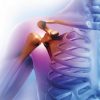
Cardiology
Cardiology polyclinic examinations
6 days of a week (except Sundays) examinations are performed between 09:00 a.m. –17:30 p.m. by specialist, associate professor, assistant professor and professor physicians.
Electrocardiography (ECG)
It is recording of electrical functions created in the heart, in order to examine the working of neural conducting systems of the heart muscle. ECG is helpful for the diagnosis of heart diseases ehen it is evaluated together with the findings and dysfunctions detected by the doctor during examination and other tests and graphs were also taken into consideration. It has an indispensable importance especially for the diagnosis of cardiovascular obstructions, rhythm disorders, heart valve disorders and heart failure.
STRESS EFFORT (Treadmill) Test
Stress effort test is a test applied for to evaluate the presence of cardiovascular diseases, determination of the effectiveness of treatment of a known cardiovascular disease, detecting whether irregularity in the heartbeats, in other words arrhythmias are occurring or not during effort, to examine the effort capacity of the patient in various heart diseases, to test the effect of effort on blood pressure in patients with hypertension and to determine the operation time in valvular heart diseases. During effort stress test, the patient walks on a treadmill. Speed and the tilt of the walking is adjusted by doctor and increase of heart beats are provided. Treadmill test is a test which is performed by serial ECG recordings and measurements of blood pressures technique while the patient is walking on treadmill with certain speeds. It provides to detect the abnormal findings after effort which cannot be detected during resting ECG. Stress effort test requires a specialist doctor from beginning to the end. Stress effort test provides great convenience for the early diagnosis and treatment of the cardiovascular diseases. Its result is given just after the test.
While coming to Stress (Treadmill) test appointment;
• Eat a light meal 3 hours before the test, then afterwards you can drink a few amount of water if you need however do not eat.
• Please bring your previous ECG’s along with you.
• Male patients should have chest shaving.
• Female patients better to wear two pieces of clothes.
• Your doctor may ask you to stop some of your medications before the stress test. Please ask this to your doctor.
Transthoracic Echocardiography (ECHO)
Echocardiography is the examination of heart structure and performance via sound waves (ultrasound). Since it is a method without including radiation and there isn’t any medication used during the procedure, it can be applied to everyone including pregnant women and newborn babies without any risk and pain. The patient is asked to lie on a stretcher during the procedure. The equipment which has insulator property and water-based gels spread on it called probe which sends sound waves is put on the chest region by doctor in various positions and allows to display the heart. Very detailed measurements and analyses were performed on these images. Diagnosis of the all structural heart diseases (heart enlargement, cardiac muscular diseases, valvular heart diseases, hypertensive heart diseases, heart failure, pericardium diseases, intra cardiac masses, clots, heart tumors, congenital heart abnormalities, diseases of the largest diameter vessel in the body called aorta) are made with echocardiography. There isn’t any preparation needed for echocardiography. However, it is appropriate to wear a comfortable dressing while coming to your echocardiography appointment. Also, a request document from the doctor of the patient who decided to the procedure including what purpose the procedure will be done and patient’s medical information should be obtained and should be delivered to the doctor who will perform echocardiography. Echocardiography is a routine method. You don’t need a specific preparation while coming to the appointment. Its result is given just after the test.
Three Dimension Trans thoracic Echocardiography
Medipol Mega Hospital Complexes have the world’s most developed echocardiography equipment. Three dimensional echocardiographs are used for valvular heart diseases and heart failure in cases where two dimensional echocardiographs are insufficient and provide valuable information. Its result is given just after the test.
Stress Echocardiography
Stress echocardiography is used for to examine whether there is an occlusion or contraction or not in the coronary arteries supplying heart, to determine whether a treatment except than medical treatment is needed or not in patients who had heart attack ( myocardial infarction) and grading the severity of valvular heart disease. It is an easy, reliable technique providing very important information.
Stress echocardiography is simply performed by examination of the heart with sound waves before and after the “created stress”. There isn’t any pain felt during this procedure. Stress method going to be used is to have effort on treadmill equipment, in other words increasing heart workload by providing walking with tempo. Stress echocardiography is also performed by giving medication from arm veins too. Its result is given just after the test.
Before stress echocardiography procedure
• Please come to your appointment with 4 hours of fasting
• Please consult with your doctor whether you will stop or not your medications before the procedure
• Please bring comfortable pair of shoes and clothes while coming to the procedure
• Please bring previous tets related with heart along with you
After the test
• Please do not eat for 2 hours
• Please do not drive since sleeping and dizziness will continue for a few hours
3 dimensional Trans esophageal Echocardiography (TEE) Test
With this method which is available only a few centers in our country, congenital heart holes, leakages of the valves in patients with prosthetic heart valves and detection of other diseases which is difficult to be detected with 2 dimensional TEE are provided. Things needed to be done before and after are same with the abovementioned information. Its result is given just after the test.
Rhythm (ECG) Holter
ECG Holter is a device which can be attached to the belt like cell phone. It is attached to the chest via 3-4 cables and electrodes (made of soft plastic, 3-4 cm in diameter adhesive material). While the patient continued to normal daily life, the equipment records the heart electro during the planned time. At the end of the time, the device is removed and the records are analyzed with the computer. With this equipment; all the rhythm disorders originated from the heart like short duration palpitations which cannot be seen during the examination, chest pains and fatigue can be detected. It should be consulted with the doctor whether the medications will be stopped or not before Holter test. After the Holter device is attached, especially the events creating the complaints should be repeated like coffee consumption, climbing stairs etc.
Event Recorder
These devices are like ECG Holter devices and used especially for the detection of rhythm disorders which are not frequently repeated. Device can remain on patient up to 14 days. It has the property of recording only when there is a complaint (recording duration can be adjusted as much as wanted).
Tension Holter
It is recording of the blood pressure and pulse by measuring the patient’s blood pressure with frequent intervals whole day. In the measurements performed between 24-72 hours and early diagnosis can be made in patients without previous hypertension. The patients’ blood pressures are measured for frequent intervals and blood pressures and pulse rates were recorded during their daily activities, sleep, and during rest. By this, treatment is arranged by determining in which hours of the day there are high blood pressures in patients with long term hypertension. The patients without previous hypertension are diagnosed early and helps to direct them for treatment.
Tilt Table Test
It is a test applied for the diagnosis of fainting (syncope) developed with sudden decrease of blood pressure and/or heart rate due to remaining still standing for long periods or sudden standings after long term sitting. It is used for the differential diagnosis of syncope. Tilt Table test is performed in the polyclinic on a table that can be tilted. The patient lie on the table and then the table brought to upright position. Excessive decrease of blood pressure and/or heart rate shows abnormal response.
Interventional procedures performed in Medipol Mega Hospitals Complex Heart Health Center
Temporary Pacemakers
In case of extremely slow heartbeat due to inability of the stimulation centers to generate enough speed stimulations or failure to deliver the created stimulus to sub-centers, pacemakers needed which are placed to the body for to maintain the heartbeat to conduct the patient’s normal life. The procedure is generally performed under local anesthesia by placing the thin wires called electrodes inside the heart through large veins at neck, chest or inguinal and attachment of this to a generator outside the body. This procedure can either be performed at bedside or under scope. The procedure generally lasts for 20-30 minutes. When the temporary pacemaker requirement is eliminated, the wire placed inside the heart is removed out.
Pacemakers
Millions of people in the world carries pacemaker. These advanced technology product small devices are used for many cases; from preventing the slowing of the heart rate to treatment of heart failure, to the task of heart pumping to prevention of sudden deaths. The device which eliminates the complaints after inserted; increases the life quality of the patient and helps to returning back to normal life. Basically there are 3 types of pacemakers: Single and double wired pacemakers that prevents the decreasing of heart rate, 3 wired pacemakers used for treatment of heart failure and defibrillators that saves the lives by giving electroshock in case of not performing pumping function due to very high rhythm. Pacemakers are inserted to the people who have heart rhythm disorders and cannot continue their lives normally. These patients are able to return back to their normal lives with pacemakers. They can return back to work, can perform house duties, drive, travel, and swim, and continue to their hobbies and sexual lives. People with pacemakers should carry their pacemaker ID along with them at all the times. They should learn the closest clinics when they travel. The performance of the pacemaker must be monitored after inserted. Pacemaker which is a small computer as well can be read from outside with a method called telemetric method by the aid of an outside computer. By this, the information is reached such as; how was the patient’s heart rate, how long has the pacemaker worked, were there any own rhythm time to time or attached to the pacemaker at all the time, were there any other rhythm disorders occurred. Also it is possible to program the pacemaker from outside such as the voltage of the pacemaker or the values needed to keep the heart rate. The patient, during the 7 years of the average lifetime of the pacemaker should have to come to controls in every 6 months. Since the pacemaker run out can be detected previously, these controls are very important.
Electrophysiological study
Electrophysiological study is a diagnostic and treatment method performed in electrophysiology/angiography laboratory by placing thin wires named electrode catheter inside heart, passing through thin sheaths placed inside inguinal veins. Electrical signals obtained directly inside the heart are evaluated through developed computers and deviations from normality are investigated. By this, it can be understood whether the main central stimulation system is working properly or not and whether the conducting system is performing its function safely or not. In most cases, in patients with palpitation complaints as excessive heart beats, the fast heart beats the patients are complained are created by the stimulations given through these wires with specific methods and causes of occurrence are investigated. When the presences of short circuits are detected, a specific current of point energy formed with radio waves is applied and tachycardia can be treated completely. By this method, permanent treatment of the most of the fast heart rates (tachycardia) became possible currently. Diagnostic electrophysiological examinations last for 30-60 minutes. If a treatment intervention is required this can be a procedure lasting for 1-4 hours.
Catheter Ablation
Catheter Ablation is the treatment of rhythm disorders by giving radio waves. This method is used in rhythm disorders which cannot be controlled with medications or in case that the patients do not want to take lifelong medications. In some cases rhythm disorders can be of life threatening importance. In such cases, catheter ablation may be required directly. Basicly the procedure is performed by anesthetize the needle entrance points with local anesthesia and in some cases under general anesthesia. Sedative medications can be applied during the procedure to make the patient feel comfortable. Success rate of the treatment of tachycardia rhythm disorders with catheter ablation varies between 70-100% according to the type of the palpitation targeted to treat and placement of the short circuit. Success means is the treatment which palpitation never occurs again. Recurrence of the palpitation after successful procedure varies according to the type of the rhythm disorder.
For example this probability is between 3-5% in palpitations due to short circuits in heart. In Medipol Mega Hospitals Complex, besides the radiofrequency ablation method called “cautery’’, arrhythmia treatment is performed with cryo-ablation method called as “freezing’’ is performed as well.
Mitral Balloon (Valvuloplasty)
Mitral stenosis; is a disease that gives symptoms in advanced stages due to the involvement of the heart valves by the childhood disease “Acute Rheumatic Fever”. Mitral stenosis is narrowing that makes the blood difficult to pass from one chamber of the heart to another. For this reason, blood accumulates in the lungs as fluid. And this causes the patient to feel dyspnea. Medical treatment is sufficient in mild stenosis however Mitral Valvuloplasty or open heart operation is performed in moderate and severe stenosis. Mitral valvuloplasty is an interventional procedure performed by entering from inguinal with a catheter, switched to the left atrium from the right atrium of the heart by intervening veil pierced with a special needle sent through a sheath. The needle is removed from the sheath and guide wire advanced to left atrium. The movements of the wire are monitored in the screen. After the wire is placed to the correct place, the balloon is advanced on the wire and placed inside the narrowed valve. The balloon is inflated where the valve is narrow and by this the valve is enlarged as much as possible. When performed in appropriate patients, the success rates of the mitral balloon treatment is as much as successful as in patients who have undergone successful heart surgery.
Advantages of Mitral Valvuloplasty
• The patient is conscious during the procedure since it is performed under local anesthesia. Right or left inguinal region is anesthetized and the balloon is advanced to heart through a small hole opened here.
• By this, the necessity of opening chest cage, stopping heart and usage of heart-lung machine are eliminated
• The patients are followed in service instead of intensive care unit after the procedure
• The patients can stand the next day
• Most of the patients can be discharged the next day
• In patients that the valve is opened with Mitral balloon and do not have arrhythmia, blood thinning medications are not required after the procedure.
• Decline is recorded in the complaints of the 90% patients with balloon mitral valvuloplasty. This recovery can last up to 20 years; most of the patients experience relief for 5-10 years at least.
Catheter Ablation
Catheter Ablation is the treatment of rhythm disorders by giving radio waves. This method is used in rhythm disorders which cannot be controlled with medications or in case that the patients do not want to take lifelong medications. In some cases rhythm disorders can be of life threatening importance. In such cases, catheter ablation may be required directly. Basicly the procedure is performed by anesthetize the needle entrance points with local anesthesia and in some cases under general anesthesia. Sedative medications can be applied during the procedure to make the patient feel comfortable. Success rate of the treatment of tachycardia rhythm disorders with catheter ablation varies between 70-100% according to the type of the palpitation targeted to treat and placement of the short circuit. Success means is the treatment which palpitation never occurs again. Recurrence of the palpitation after successful procedure varies according to the type of the rhythm disorder.
For example this probability is between 3-5% in palpitations due to short circuits in heart. In Medipol Mega Hospitals Complex, besides the radiofrequency ablation method called “cautery’’, arrhythmia treatment is performed with cryo-ablation method called as “freezing’’ is performed as well.
Mitral Balloon (Valvuloplasty)
Mitral stenosis; is a disease that gives symptoms in advanced stages due to the involvement of the heart valves by the childhood disease “Acute Rheumatic Fever”. Mitral stenosis is narrowing that makes the blood difficult to pass from one chamber of the heart to another. For this reason, blood accumulates in the lungs as fluid. And this causes the patient to feel dyspnea. Medical treatment is sufficient in mild stenosis however Mitral Valvuloplasty or open heart operation is performed in moderate and severe stenosis. Mitral valvuloplasty is an interventional procedure performed by entering from inguinal with a catheter, switched to the left atrium from the right atrium of the heart by intervening veil pierced with a special needle sent through a sheath. The needle is removed from the sheath and guide wire advanced to left atrium. The movements of the wire are monitored in the screen. After the wire is placed to the correct place, the balloon is advanced on the wire and placed inside the narrowed valve. The balloon is inflated where the valve is narrow and by this the valve is enlarged as much as possible. When performed in appropriate patients, the success rates of the mitral balloon treatment is as much as successful as in patients who have undergone successful heart surgery./p>
Advantages of Mitral Valvuloplasty
• The patient is conscious during the procedure since it is performed under local anesthesia. Right or left inguinal region is anesthetized and the balloon is advanced to heart through a small hole opened here.
• By this, the necessity of opening chest cage, stopping heart and usage of heart-lung machine are eliminated
• The patients are followed in service instead of intensive care unit after the procedure
• The patients can stand the next day
• Most of the patients can be discharged the next day
• In patients that the valve is opened with Mitral balloon and do not have arrhythmia, blood thinning medications are not required after the procedure.
• Decline is recorded in the complaints of the 90% patients with balloon mitral valvuloplasty. This recovery can last up to 20 years; most of the patients experience relief for 5-10 years at least.
Coronary Angiography
Coronary angiography is a method used for the diagnosis of the determination of coronary arteries that supply heart. They develop due to cardiovascular diseases. Coronary angiography detects which part and how much of the coronary arteries are narrowed or obstructed. By determining the narrowing and obstructions of the coronary arteries provides the direction of the treatment as required. Inguinal or arm arteries are used as intervention site for coronary angiography. First the sheath is placed to the artery at intervention site and via this sheath, by using various catheters, and artery structures are observed by giving opaque agent to the beginning of the coronary arteries. Coronary angiography is performed is specifically deployed angio halls. After the intervention is terminated, the sheath placed to the intervention site artery is removed. Pressure is applied to that site and stopping of bleeding is provided. After performing a tight bandage, the patient is taken to the bed. Coronary angiography is completed after 20 or 30 minutes the patient has been taken to angio room. This time can be long in some cases (bypass patients, patients who had previous heart operations and patients with obstruction in inguinal or arm arteries etc.). The patient is needed to be hospitalized for coronary angiography. The patient is rested for 6 hours after the procedure is completed and then standing up is provided can be discharged if general status is appropriate and the doctor is confirmed. In some cases suture system may be used after the sheath is removed. These patients can stand up earlier and discharged. One should be contacted to angio secretary few days ago before coming to angiography and information must be obtained about what to do.
Cardiac Catheterization
Cardiac catheterization is a method performed similar to coronary angiography procedure by using inguinal or arm arteries however mostly requires simultaneous venous intervention too. Cardiac catheterization is a diagnostic method used for the diagnosis and whether a different treatment is necessary or not for congenital or acquired diseases related with the structure of heart, abnormalities, congenital or acquired heart holes. Cardiac catheterization procedure generally lasts for approximately 30 minutes. After the procedure, sheaths placed to artery and veins are removed and stopping of the bleeding is provided by outside compression. Tight bandage is applied to that region and the patient is taken to bed. Generally 6 hours of rest after the procedure is sufficient. At the end of the rest, the patient can be discharged if the doctor finds appropriate. Life risk is very low in cardiac catheterization and coronary angiography. One should be contacted to angio secretary few days ago before coming to cardiac catheterization and information must be obtained about what to do.
Opening of chronic totally occluded arteries
Some of the arteries are remained 100% occluded in some of the patients who had heart attack and the patients mostly require operation in order to open the arteries. Opening of the 100% occluded coronary arteries without need of operation with balloon and stent procedure is performed only in significant heart centers in Turkey can be performed by the cardiologists of the Medipol Mega Hospital Complex Cardiology Department. Our clinic is consisted of the most experienced and highest number of patients about this subject.
Non-surgical Treatment of Aortic Stenosis (TAVI)
The method shortly called as TAVI (Trans catheter Aortic Valve Implantation); in fact explains the procedure of setting aortic valve by using catheter method without performing open heart surgery. Biologic heart valves are used during this procedure which are used in the valve changing operations both in world and in our country. In TAVI method, this biologic valve is placed inside a stent sheath and when the stent is opened the valve is tightly hold and placed to the region. 2 different techniques can be used during this method: In the first technique, the valve is advanced with the aid of catheter from inguinal to heart like in the angiography applications and placed here bt opening the stent mechanism. Other technique is applied if there is an occlusion in the arteries at inguinal or abdominal region that will be used to reach to the heart. In this technique, approximately 4-5 cm small incision is performed at chest anterior wall and reached to the end portion of the heart and valve is placed via the catheter advanced from here. In both methods, stopping heart and open operation are not required. The procedure can be performed under local anesthesia without general anesthesia. The patients are taken to coronary intensive care unit after the TAVI procedure. A blood thinning medication is given to the patient at the moment and the patient is followed in the hospital for 4-5 days under normal patient conditions. The patient is discharged at the end of this period. The patient who has sent to home is coming back to control after a few days of rest and continue to normal life.
TAVI method is primarily recommended for high risk patients who cannot handle open surgery for valve placement. Except this, this method can be applied for the patients who have a difficulty for open surgery. For this subject, especially very old, patients with lung, liver or renal functional disorders or patients with previous open heart surgery history are accepted as high risk patients for open surgery. TAVI method is known to be effective for these patients who are not appropriate for open surgery in terms of extending life span and recovery of the patient’s clinic status.
Non-surgical treatment in heart holes
In the past, while intra cardiac holes were closed majorly with surgery, recently non-surgical closing methods are preferred operational. In patients with congenital cardiac holes, heart cannot continue normal functioning and clean and dirty bloods are mixed to each other. In our clinic, most of the holes in these patients are closed non-surgically by entering from inguinal artery, closed with a device after passing the region where the hole is with the aid of a catheter. Our patients are discharged within 48 hours after the procedure.
Non-surgical vessel repair
Endovascular method is primarily convenient for the high risk patients for open surgery. However, this method cannot be applied to every patient because the patient’s vessel structure must be sufficient for the treatment of aneurysms with endovascular method. Also, it is commonly applied in ruptured aneurysms that require emergency surgery in terms of its fast and practical applicability.The procedure is performed with local anesthesia. However, it requires to be performed under the conditions that catheter techniques can be applied and operational conditions are present. For this reason, providing the environment which is called hybrid operating room, where both operation room conditions provided and catheter treatments are performed and advanced imaging methods can be applied, is a big advantage.
EVAR: In endovascular treatment method, aorta region that the aneurysm formed in the abdominal vessels is treated with a stent that is advanced with the catheter from inguinal regions.
TEVAR: In endovascular treatment method, aorta region that the aneurysm formed in the chest vessels is treated with a stent that is advanced with the catheter from inguinal regions.
A new treatment method in resistant hypertension: Renal Denervation
There are nerves called “sympathetic” around the renal vessels that cause elevation of blood pressure. This sympathetic system is cauterized via a specific material inside the vessels with a method like angio without anesthesia. This method which depends on the cautery of sympathetic nerves causing hypertension is called as renal sympathetic denervation. This method can effectively treat the persistent tension which is not decreased or recurred after a while after decreasing and affecting the patients’ quality of life severely. 80-90% of success provided by renal denervation method in persistent hypertension treatment provides decrease the number of the blood pressure medications of the patients and controlling blood pressure.
Procedures performed in Medipol Mega Hospital Complex Nuclear Medicine Department
Myocardium Perfusion Scintigraphy (Thallium Test)
Blood supply of the myocardium during rest or under stress is examined with this test. It is examined whether there is an obstruction or not in the vessels supplying heart and if there is, whether this obstruction creates a dysfunction of the supplying of heart or not. It is a more sensitive test than stress effort test. First, the patient is given intravenous radioactive material and resting images are obtained in the device called gamma camera, then the patient is walked on the treadmill or stress test with medication is performed, again radioactive agent is injected. Images are obtained in gamma camera again. Radioactive material reaches to heart and myocardium. This material is less involved at the places where there is atherosclerosis. Gama camera is the name of the device provides taking images. It turns around the heart with certain periods and transmits the radioactive signals from the heart to a computer. The computer transforms the obtained signals to a colored image and performs the blood supply analysis of the all regions of the heart.
In which cases myocardium perfusion scintigarphy is requested; In the determination of the chest pains relationship with heart disease, in the diagnosis of cardiovascular diseases, determination of the vitality of myocardium after myocardial infarction, in the determination of the procedure’s benefit and necessity before and after coronary balloon angioplasty, as a disease screening test in patients who have risk factors of cardiovascular diseases, detecting the importance of the vascular disease in people with cardiovascular disease, detecting the effectiveness of treatment after coronary bypass surgery or medical treatment, to detect whether the patients had or not myocardial infarction and for the determination of the wideness and severity of the myocardial infarction.
Procedures performed in Medipol Mega Hospital Complex Radiology Department
Coronary CT Angiography
Computerized Tomography (CT) Coronary Angiography can be performed with multi slice “Multi Detector –multi slice-CT” device without requiring preliminary preparation, without catheter and only within a breath holding time period. CT Coronary Angiography is a new groundbreaking method in imaging area which can be performed bloodless and painless and can be performed faster and easier than classic angiography. It eliminates the necessity of hospitalization. The patient continues to daily life after the procedure. With this system, coronary angiography of the people with risk factors can be easily done. It has been observed that, especially the patients who had previous classic angiography prefer this method and with CT angiography many patients are intervened and treated earlier. 256-detectores CT device which is the most developed CT Coronary Angiography device in Turkey is available in Medipol Mega Hospitals Complex.
Cardiac MR
It is an easy and non-medication or non-procedure requiring imaging method that reveals the diseases of pericardium, heart chambers, heart valves, myocardium, structures of the large arteries exit from the heart, and some coronary diseases (for now only the main branches). The duration of procedure is approximately 45 minutes and does not require any preliminary preparation.