Description
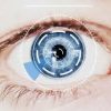
ABOUT THE RETINA
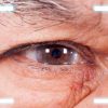
he retina is a layer of the eyeball lining its back wall, which consists of light-sensitive cells. Retinal diseases threaten our sight directly.
Retinal Diseases
• Hemorrhages related to diabetes and hypertension
• Retinal vein occlusions
• Retinal detachment / tears
• Yellow spot disease
• Congenital retinal diseases
• Fluid retention under the retina, retinal edema
• Foreign bodies in the eye
• Macular holes
• Vitreoretinal surface disorders
• Retinal tumors
Symptoms of Retinal Disease
• Sudden or gradual loss of vision
• Distorted vision
• Flashes
• Eye floaters
• Curtained vision
• Temporary, short-term loss of vision
• Dark areas in the field of vision
Negative Effects of Diabetes and Hypertension on the Retina
Diabetes and hypertension have adverse effects on all the body’s systems and their first and most important adverse effect occurs in the eyes. Dilatation of the retinal vessels and permeability impairments occur as a result of these diseases.
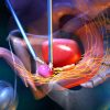
Treatment of Retinal Diseases at Dünyagöz
Early diagnosis, preventive medical therapy and undelayed accurate surgical intervention is of vital importance in retinal diseases. At Dünyagöz, retinal surgeries are performed by a team which has been specially trained for intraocular microsurgeries, using a comprehensive set of instruments and devices.
RETINA DECOLLEMENT
Retinal detachment develops due to tears or holes in the retina. It is frequently seen in patients with high myopia. It may develop at any age but more in middle-aged and elderly people.
The retinal layer becomes strained as the eye’s anteroposterior diameter increases and the strained area starts to thin and break down. Sporadic thinning and breaking down can occur around the retina in some familial or degenerative diseases and infections too. Meanwhile, the vitreous gel starts to lose its homogeneity and deteriorate for the same reasons; the gel’s consistency changes and it slowly separates from the retina. This separation is called vitreous detachment. The vitreous tissue shrinks and becomes opaque at places; as it passes the eye’s visual axis, the person perceives this as eye floaters or a curtain of smoke. Retinal detachment may lead to partial or total loss of vision if not treated without delay by a doctor who is experienced in retinal diseases.
Causes and Symptoms of Retinal Detachment
If untreated, retinal diseases may result in permanent blindness. Symptoms such as glare, floaters or sudden loss of vision may indicate a significant eye disease like retinal detachment. Results which may lead to loss of vision can be prevented through early diagnosis, detailed examination and a timely – and most important of all, accurate – treatment. Retinal surgeries are major operations which require important sterilization measures and use of high technology and otherwise may result in loss of vision.
When the macula (the eye’s visual center) separates from the tissue underneath, central vision is lost. In long-term detachment, intraocular balances are disrupted and the eyeball starts to get smaller. Sudden, severe or perforating trauma to the eye can be a cause of detachment. In diabetes and some degenerative diseases, strands are formed in the vitreous that pull away at the retina, leading to traction-related detachment. Though rare, detachments may develop without any tearing in the eye, as in certain infections, tumors and especially hypertensive crises that occur during pregnancy.
DIABETIC RETINOPATHY
________________________________________
The most common cause of diabetes-related blindness is “Diabetic Retinopathy”.
Diabetes is a metabolic disease characterized by elevated blood glucose as a result of insufficient release or action of insulin.
Diabetes particularly affects the eye’s neural layer (retina) and the capillary vessels in this layer, disrupting their function and leading to loss of vision. Retinal disorders associated with diabetes are called diabetic retinopathy.
In diabetic patients, an eye examination should be performed from adolescence on in young people and at the time of diagnosis in individuals that develop diabetes after 30 years of age. If their retina is normal, an annual eye examination should be performed in diabetic patients. Once a retinopathy starts, the interval of check-ups can be reduced to 3-4 months.
Causes of Diabetic Retinopathy
The primary risk factor for diabetic retinopathy is the duration of diabetes. The incidence of retinopathy increases particularly after the 10th year following the diagnosis of diabetes. In young patients with type 1 or insulin-dependent diabetes, the incidence of retinopathy increases with age after adolescence.
Keeping blood glucose under control is an important factor. Unstable blood glucose levels and sudden increases and decreases in blood glucose make retinal deterioration and disease progress easier. Pregnancy, hypertension, elevated blood fats (hyperlipidemia) and kidney disease are factors aggravating retinopathy.
Diabetes damages retinal capillary vessels, causes cell loss, disrupts vessel permeability and causes retention of fluid and fatty substances in the macular area, hence obstruction of capillaries, leading to areas with no blood supply. New vessels are formed in the retina which can bleed spontaneously. Bleeding occurring in front of and inside the retina may leak into the eye’s posterior chamber. Venous membranes are formed in the retina, leading to serious losses of vision and painful elevations in eye tension.
MACULAR DEGENERATION (YELLOW SPOT)
________________________________________
Yellow spot disease (macular degeneration) is a retinal disease that affects
central vision. It is quite common after the age of 55 and may lead to loss of vision in the event of progression.
Who does yellow spot disease affect?
• People older than 55 years of age
• Those with hereditary risk
• Smokers
Risk Factors
The main risk factors for age-related yellow spot disease include the person’s age and hereditary characteristics. Other risk factors are hypertension, smoking, nutrition, elevated lipid-cholesterol levels, long-term exposure to sunlight and being overweight.
Symptoms of the Disease
• Loss of vision
• Wavy or broken view of objects/lines
• Shadows in front of the eye
• Impaired visual quality
• Trouble discerning colors
Age-related and hereditary factors are impossible to eliminate. However, other risk factors can be controlled. If there is hypertension, it can be controlled. If smoking, the patient should quit. Filter sunglasses should be worn for sun protection. A Mediterranean diet is recommended. It is recommended to avoid butter, red meat and cholesterol-containing food.
How many types of yellow spot disease are there and what are their results?
There are two types of yellow spot disease; dry and wet. The dry type accounts for 90% and the wet type accounts for 10% of all cases. However, early diagnosis is more important in the wet type since it leads to loss of vision. Progressing more rapidly than the dry type, this disease leads to sudden loss of vision accompanied by impaired color vision and a loss in contrast sensitivity, and to blindness because of bleeding in the new vessels that are formed in the retina and macula over time.
Treatment of Yellow Spot Disease
Preventive treatment is given in the dry type of yellow spot disease; intraocular injection and photodynamic treatment is administered in the wet type. Antioxidant vitamins A, C and E, lutein and zinc are used in preventive therapy. Intraocular injections are made by numbing the eye with an eye drop. The patient feels no pain during this procedure. In photodynamic therapy, a drug called verteporfin is first administered intravenously in a special combination and a low power laser is then applied.
Drug Injection in the Treatment of Yellow Spot Disease
A kind of protein (anti-VEGF antibody) is used for intraocular injection therapy. FDA-approved anti-VEGF drugs are injected into the eye with a needle for treatment of the wet type of yellow spot disease, which occurs in one in every three people aged 75-85 years. The anti-VEGF drug used during the treatment inhibits the protein which is released by the eye cells behind the eye in the event of disease and forms new vessels, thus preventing loss of vision. Injected into the eye at 4-6 week intervals, the drug stops the development of new vessels in the yellow spot and improves the patient’s complaints significantly. The injection is made at least 3 times but some patients require more. Injection intervals vary between 4-6 weeks depending on the patient’s response to therapy.
If untreated, yellow spot disease results in 95% loss of vision and ultimately in total loss of vision. Vision is reduced to a level which is legally accepted as blindness and patients become unable to see what they are looking at. For instance, the patient cannot see the face of the person in front of them, but can see their
arm or leg. These patients usually cannot leave their house alone; they can do their house chores by themselves but need the help of others in many tasks. Since they can’t see, they can’t read, write, watch TV or drive.
Amsler Grid Test in Yellow Spot Disease
This test is not the equivalent of a routine eye exam. However, it is a graph that you can self-administer to identify the early symptoms of yellow spot disease. Experts recommend that everyone older than 40 years of age take this test.
GRAPH
Method of Administering the Amsler Grid Test
• Wear your eyeglasses or contact lenses if you use a pair.
• In a well-lit room, hold the above graph approximately 30-40 cm away from your face in a fixed way.
• Cover one eye with your hand and focus on the dot in the middle. You should be able to see all 4 corners of the large square in the
graph.
• Test your other eye in the same way.
• You could have symptoms of yellow spot disease if the lines appear wavy, broken or blurry or if you can’t see the corners.
In that case, you should see an ophthalmologist specialized in retinal diseases as soon as possible.
ETINOPATHY OF PREMATURITY (ROP)
________________________________________
What is retinopathy of prematurity?
Retinopathy of prematurity is described as one of the most important eye health issues affecting premature babies. Vessels in the eyes of infants continue to develop until the time of birth. In premature babies, such development continues after birth since it is not completed yet. High concentrations of oxygen given to keep premature babies alive cause the vessels in the eyes to develop abnormally. As a result, retinopathy of prematurity (shortly called ROP) develops in the retinas of infants whose vascularization is not complete. Unless treated at an early period, this leads to blindness in both eyes. Therefore, premature babies should be examined by an ophthalmologist.
Which babies does retinopathy of prematurity affect most?
A normal pregnancy lasts 40 weeks or 280 days. If delivery takes place before week 37 is completed, the baby is considered to be premature. Infants born weighing less than 2,500 grams are termed “low birthweight” babies. Two thirds of these infants are premature babies.
Retinopathy of prematurity is most common in those whose birthweight is less than 1000 grams. Therefore, all infants born weighing less than 1500 grams and earlier than week 32 should be examined for ROP. Early diagnosis and treatment of ROP in newborns is possible through the collaboration of specialist pediatricians and ophthalmologists. Pulmonary and coronary disorders, severe infections and any brain-related problems occurring in infants also increase the risk of retinopathy. Treatment with early diagnosis is possible; delayed treatment results in blindness in both eyes.
When should babies have their eye examination?
An eye examination should be performed 4-6 weeks after birth. ROP has five stages from mild to severe and success is correlated with disease stage. Follow-up is sufficient in the first two stages; from the third stage on laser and cryo treatment should be started because the best results are achieved in the third stage. In the fourth and fifth stages, the result of the necessary surgical intervention is not successful. Performing an eye examination in the first month following birth is important for all newborns since timely diagnosis and successful treatment is made possible not only for ROP but also for many eye diseases including high eye pressure, lazy eye, lacrimal duct occlusion and strabismus.
Pediatric Anesthesia
FDA (American Food and Drug Administration)-approved technologies and materials are used in the monitoring and treatment of pediatric eye diseases. If anesthesia is required, it is administered by anesthesiologists specialized in pediatric anesthesia. Anesthesia is given in sterile operating rooms, on operating tables designed to fit pediatric patients and using personal, single-use medical supplies.