Description
Brain, Nerve and Spinal Cord Surgery
Surgical Treatment of Congenital Diseases of the Brain
Arnold- Chiari Malformation
Arachnoid Cysts
Dandy-Walker Malformation
Hydrocephalus
Surgical Treatment of Brain Tumors
Surgical Treatment of Benign Tumors of the Brain –
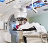
Surgical Treatment of Malignant Tumors of the Brain
Tumors Originating from Brain Tissue
Tumors Developing After Metastasis to the Brain
Surgical Treatment of Vascular Diseases of the Brain
Surgical Treatment of Cerebrovascular Bubbles (Aneurysm Surgery)
Surgical Treatment of Cerebrovascular Tangles (AVM Surgery)
Surgical Treatment of Intracerebral Hemorrhage
Surgical Treatment of Brain and Neck Vascular Occlusion
Carotid Endarterectomy
Bypass Surgery
Surgical Treatment for Re-bleeding
Decompression Surgery in Brain Infarction
Surgical Treatment of Brain Injuries
Emergency Surgical Treatment of Brain Injuries
Skull Crashes
Injury-Related Brain Hemorrhage
Late Surgical Treatment of Brain Injuries
Surgical Treatment of Chronic Subdural Hematomas
Surgical Treatment of Brain Function Disorders
Surgical Treatment of Movement Disorders
Surgical Treatment of Sara’s Disease
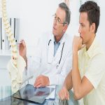
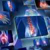
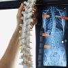
Spinal Surgery
Surgical Treatment of Congenital Diseases of Spine and Spinal Cord
Surgical Treatment of Tumors of the Spine and Spinal Cord
Surgical Treatment of Vascular Diseases of the Spinal Cord
Surgical Treatment of Lumbar Hernia
Microdiscectomy
Endoscopic Discectomy
Surgical Treatment of Neck Hernia
Surgical Treatment of Lumbar Slips
Surgical Treatment of Spinal Canal Stenosis
Surgical Treatment of Spine and Spinal Cord Injuries
Surgical Treatment of Spinal Disorders
Nerve Surgery (Nerve tumors and all nerve syndromes)
Surgical Treatment of Nerve Tumors
Surgical Treatment of Nerve Injuries
Surgical Treatment of Nerve Congestion
Thoracic OutLet Syndrome (Cervical Costa)
Carpal Tunnel Syndrome
Ulnar Gutter Syndrome
Peroneal Nerve Entrapment Syndrome
Tarsal Tunnel Syndrome
Brain, Spinal Cord and Nerve Surgery in Childhood Diseases (Pediatric Neurosurgery) Meningiomas
They make up about 15% of all brain tumors. They
originate from the membranes that surround the brain. They are usually
slow-growing, benign tumors. They can be located on the outer surface of
the brain, as well as in the base of the skull or deep within the
brain. Although they can cause complaints in patients depending on where
they are located, headache is the most common complaint. Following this,
epileptic seizures, loss of strength or sensation in the arms or legs, visual
impairment, etc. There may also be complaints. If they can be
completely removed (which is sometimes not possible), they are less likely to
recur.
Pituitary Adenomas
They constitute 10-15% of all brain tumors. They
originate from the pituitary gland located under the brain, which is connected
to the brain by a stalk. Although they are mostly benign tumors, they may
tend to grow again despite being removed. There are types that can secrete
hormones as well as types that do not secrete hormones. Those who secrete
hormones cause different complaints in patients according to the type of
hormone they secrete. For example; Patients with prolactin secreting
prolactinomas have complaints such as menstrual irregularity, milk coming from
breasts or decreased sexual power, while tumors secreting growth hormone cause
acromegaly or gigantism. In patients, there is growth in structures such
as hand, foot and facial bones, and external physical properties can change, as
well as internal organs, diabetes (Diabetes Mellitus), heart failure,
etc. they can cause. Again, in patients with ACTH hormone secretion,
the disease we call Cushing’s disease appears in patients, and patients have a
face, obesity, purple lines in the abdomen, muscle wasting and weakness,
osteoporosis, diabetes, high blood pressure, acne, increased hair growth and
baldness, susceptibility to infection, fat accumulation on the back (buffalo).
hump). Hormone-secreting types are diagnosed relatively earlier than
non-secreting types. The types that do not secrete hormones are usually
more insidious. Diagnosis can be made when vision loss occurs, mostly as a
result of pressure on the visual nerves. Headache can be felt in almost
all pituitary adenomas due to the tension in the brain membranes during the
growth phase of the tumor.
Unsuccessful Lumbar Surgery Syndrome
After lumbar hernia and lumbar surgical interventions,
many patients experience pain due to various reasons. This condition is
called failed back surgery syndrome or insufficient back surgery syndrome.
Slipped Lumbar Spine
There are two main parts of the spine. Vertebral
body (vertebral corpus) and posterior elements. The posterior elements
form a canal in the posterior part of the vertebral corpus. These channels
join together to form the spinal canal (Spinal canal). In addition, the
posterior elements connect with the lower and upper vertebrae by joints called
facet joints. There are discs between vertebral bodies. This
structure provides flexibility of the spine.
Sometimes there is an opening in the parts (pedicle)
connecting the back of the spine to the body due to congenital or acquired
reasons. This opening is called Spondylolysis. As a result of
spondylolysis, the forces applied to the vertebrae cause the upper vertebra to
slide forward on the lower vertebra. This event is called
Spondylolisthesis (slipped vertebra).
Spondylolysis and spondylolisthesis are seen in 5-6% of
the population. It is most common in the lowest two vertebrae of the
waist. Because this region is the region that is exposed to the most shear
force.
Lumbar Hernia
With the five vertebrae and the discs between these
vertebrae that act as shock absorbers, the lumbar region located on the sacrum
(rump) forms the most mobile part of the spine after the neck. The
majority of the movement in the waist is formed by the joints between the 4th
and 5th lumbar vertebrae and the 5th lumbar vertebra and the sacrum
bone. The inside of the discs between the vertebrae is a gelatinous liquid
containing approximately 70-80% water and the outer part consists of fibers of
fibrotic bands. Over time, the liquid ratio in these discs decreases, and
the disc content, which has not broken before, becomes a breakable, breakable
form. Repetitive movements, excessive strains, posture disorders and
physical activities performed in inappropriate positions cause tears in the
outer zone called annulus fibrosis, The tear starts from the inner fibers
of the annulus and extends outwards. As a result, the dehydrated, degraded
gelatinous fluid becomes herniated outward and forces the ligaments in that
area and puts pressure on the surrounding tissues. Patients suffer from
back pain from time to time during periods of rupture in the capsule of the
disc. Most of these can be cured by just bed rest without any
treatment. However, as the disease progresses further, it compresses the
nerves leading to the leg, and in this period, leg pain becomes more prominent
in patients. Nerve fibers also resemble the same electrical cables, mostly
fibers that feel like fibers closer to the surface on the outside. Deeper
ones are the fibers that make the movement. When the dislocated disc
causes irritation in the nerve coming to the leg in patients, first of all, pain
is felt in the area where that nerve bears the sensation. If the event
progresses and the fibers carrying the sensation are damaged, numbness
(numbness) occurs in that area, if the patient is still not treated at this
stage, it is inevitable that the patient will lose strength as a result of the
effect of the fibers that make the movement. It is mostly seen in young
and middle ages. In advanced ages, it is seen with waist calcification.
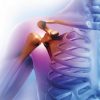
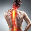
Neck Hernia and Calcification
Since cervical disc hernias and calcifications cause
almost the same complaints and are often seen together, they are described
under the same heading.
There are 7 vertebrae and 6 intervertebral discs in the neck region. There
is no disc between the base of the skull and the 1st neck vertebra and
C1-2. Vertebrae in the neck; It articulates through the discs between
the vertebrae in the front and two protrusions (facet or apophyseal joint)
between the lower and upper vertebrae in the back. The joints in the back
are called apophyseal joints. In addition, the ligaments, apophyseal
joints, apophyseal joint capsules that run in front of and behind the spinal
cord in the neck are highly affected by all kinds of diseases in the neck.
Also; Stress, occupational strains, using a
typewriter, traffic accidents, posture disorders are important factors that
disrupt neck health. The first change in the neck starts from the
discs. Initially, the water content in the discs decreases, tears occur in
the fibers on the inner side of the disc, and the gelatinous fluid in the disc
herniates from these tears and puts pressure on the nerves and soft
tissues. The herniation of the neck causes calcification in the anterior
and posterior joints of the neck and consequently loss of movement in the neck
and localized (regional) radicular (spreading) pain.
Neck Hernia and Treatment
There are 7 vertebrae on the neck. There
are cartilages between each vertebra, which serve as a cushion that we call a
disc. The condition that occurs as a result of the rupture of this
cartilage structure and the pressure on the nerves coming to the spinal cord or
arm is called cervical hernia. The patient has a severe neck pain, pain
spreading to the arm, numbness. If the cartilage, which is torn over time,
puts pressure on the nerves, weakness in the arm, and movement defects in the
whole body may occur if it also puts pressure on the spinal cord itself. Patients
who become bedridden in the very advanced stages of the disease are encountered.
Surgical treatment of cervical hernia, cervical microdiscectomy The
aim of surgical treatment is to remove the pressure on the spinal cord and
nerve tissue. Thus, the patient’s pain is relieved, and symptoms such as
numbness and weakness are provided. These attempts, made at the
appropriate time and in experienced hands, give very good results. The
only method used today is cervical microdiscectomy. In some patients,
together with microdiscectomy, a bone or synthetic prosthesis taken from the
body instead of the removed cartilage is applied.
Advantages of cervical microdiscectomy:
The least risk of tissue damage, blood loss and infection due to surgery.
To completely remove the torn cartilage under the microscope.
Postoperative pain and lack of movement limitation.
The patient’s ability to return home and work in a short time.
After the operation, the patient stands up 3 hours later and is sent home in the evening. The
patient, who needs to wear a collar for 1-3 weeks, can return to work after 10
days.
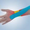
Nerve Entrapment in the Wrist Canal (Carpal Tunnel Syndrome – CTS)
The compression of the median nerve (others; ulnar nerve
little finger and half of the ring finger, radial nerve dorsum of the hand),
which is one of the three arm nerves that comes out of the neck area and
irritates the arm and hand, is called Carpal Tunnel Syndrome (CTS).
Lumbar Spondylosis
There are 5 vertebrae in the lumbar region and 5 discs
between the vertebrae. The waist area provides flexibility and mobility of
the body. It is also the place of passage of nerves that are transported
from the brain through the spinal cord and extend from there to the legs. There
are two important nerves in the lumbar region. Sciatic nerve in the back,
femoral nerve in the anterior part of the leg. The sciatic nerve extends
to the toe and is the longest and thickest nerve in the body.
The most active regions
of the waist (L4-5 and L5-S1) are the regions. For this reason, most of
the diseases related to aging and wear, such as herniated disc, calcification,
are seen in this range.
Entrapment Neuropathies of the Peroneal Nerve
The peroneal nerve consists of the posterior division of
the L4, L5, S1 and S2 roots and is separated from the sciatic nerve on the
popliteal fossa. As it descends along the outer side of the fossa, a
cutaneous branch joins with the sural nerve and the lateral cutaneous nerve
located on the anterior outer surface of the leg is separated. It rotates
around the neck of the fibula, dividing it into superficial peroneal
(musculocutaneous) and deep peroneal (anterior tibial) nerves. The
superficial peroneal nerve runs down the outer edge of the leg, innervating the
peroneus longus and brevis muscles, providing sensation to the lower anterior
aspect of the leg and most of the back of the foot.
Deep peroneal nerve descends from the anterior aspect of
the leg, before passing under the extensor retinaculum, innervation of the
tibialis anterior, extensor hallucis and digitorum longus and peroneus tertius
muscles, after crossing the retinaculum, the lateral terminal branch of the
extensor digitorum brevis muscle and the medial terminal branch of the first
and second finger on the dorsum of the foot. Provides sensory innervation of a
small area in place.
The peroneal nerve can be compressed and directly
traumatized, especially at the level of the head and neck of the
fibula. The nerve may be damaged as a result of total knee arthroplasty or
an arthroscopic procedure performed on the knee. Plasters, leg orthoses,
high boots, tight sock ties, stockings and legs may be under nerve compression
as a result of sitting for a long time by crossing their legs. In
addition, placing the patient in an inappropriate position during anesthesia
may cause compression of the nerve. In this way, compression-related paralysis
is more common in patients hospitalized for intoxication, stupor or coma.
Inversion injuries of the ankle are less common causes of
peroneal neuropathy. Acute lateral compartment syndromes can develop as a
result of athletic activity. The nerve biceps tendon can be squeezed
between the lateral head of the gastrocnemius and the head of the fibula with
the compression force created by the body weight in the muscles during
squatting. Peroneal neuropathy that develops after weight loss has also
been described. Here, nutritional deficiency, metabolic factors, or a
reduction in the protective subcutaneous tissue surrounding the nerve are
thought to cause the event and the prognosis is generally good. Although
rare, peroneal neuropathies due to tumors or cysts have been encountered. Peroneal
neuropathy is more common in diabetic patients.
In peroneal nerve lesion, signs of dorsiflexion of the
foot, eversion and weakness of the toe dorsiflexion muscle strength
accompanying the loss of sensation in the back of the foot and the anterior
lateral surface of the leg are observed. Drop foot develops in severe
lesions. The inversion of the foot is normal since the muscle that
provides the foot inversion is not innervated from the peroneal
nerve. This situation helps to make a clinical differential diagnosis
between peroneal nerve palsy and sciatic nerve or lumbosacral root
lesions. There is local sensitivity at the neck or head of the fibula.
Motor neuron disease is sometimes associated with drop
foot, but the presence of fasciculation, upper motor neuron deficits, and
preservation of the sensation distinguish motor neuron disease from peroneal
neuropathy.
Clinical deficits are different when a partial lesion
develops in the peroneal nerve. One study reported that muscles innervated
from the deep peroneal nerve tend to be more affected by muscles innervating
the superficial peroneal nerve, sometimes mistakenly referred to as deep
peroneal neuropathy.
Sometimes, half of the outer part of the extensor
digitorum brevis (EDB) muscle can also be innervated by the accessory deep
peroneal nerve, a branch of the superficial peroneal nerve. Since the EDB
is also under the volanter control of the accessory deep peroneal nerve,
complete lesion of the deep peroneal nerve may be overlooked in these patients.
The deep peroneal nerve can be compressed within the
anterior tibial compartment. In this condition called “anterior
compartment syndrome”, muscle edema causes entrapment of the deep peroneal
nerve. Edema may be caused by excessive exercise, trauma, or occlusion of
the anterior tibial artery. Decompression surgery is urgently required to
reduce neurological damage. The deep peroneal nerve can also be compressed
on the dorsum of the foot. It causes pain, paresthesia or EDB muscle weakness,
called anterior tarsal tunnel syndrome. The medial branch of the nerve can
be compressed under the extensor hallucis brevis tendon and causes only sensory
complaints at the junction of the thumb and second finger.
Superficial peroneal nerve can be held in the lateral
(peroneal) compartment due to excessive activity or trauma. Patients have
painful paresthesia complaints on the dorsum of the feet. Clinically,
local sensitivity and loss of sensation is observed approximately 10 cm above
the lateral malleolus.
L5 radiculopathy, lumbosacral plexus lesion, partial
lesions of the sciatic nerve and motor neuron disease are included in the
differential diagnosis.
It is important to stimulate the patient in order to
prevent the nerve compression in the knee area. In the vast majority of
patients, the clinical picture improves spontaneously. Surgical
intervention is indicated in unresolved cases. In the fibular tunnel, the
nerve is released. Motor function improves in 87% of cases after
decompression. Emergency intervention is required in anterior compartment
syndrome. With fasciotomy, both the muscle and the nerve are healed.