Description
Urology
Urological cancers
Prostate Cancer Prostate cancer is generally a disease of men after the age of 50. More
than 90% of men diagnosed with prostate cancer are older than 60 years of age
at the time of diagnosis. With the increase in the average age of the
population, the frequency of prostate cancer is gradually increasing in our
country. Hidden prostate cancer is present in 30% of men over the age of
70. Only some of these tumors start to grow faster at any one time and
become a dangerous disease that needs to be treated.
Causes and Risk Factors in the Formation of Prostate Cancer The causes of the disease have not been revealed until now. However, there are 3 definite risk factors revealed in prostate cancer;
Age (the risk of being seen increases with age),
Genetics (the risk is higher in those with a family,
especially a first-degree relative with prostate cancer),
Race (fairly high in black race in America).
This disease is relatively common in Europe and North America, and less frequent in East Asia. Therefore, lifestyle and living conditions are considered to be affecting the risk of getting sick. Diets
high in fat and low fibrin probably facilitate the occurrence of prostate cancer.
There may be an apparently higher risk in workplaces where the heavy metal Cadmium is present, for example in men working in the tire industry.
Close relatives of prostate cancer patients get this disease more often than other segments of the society. Accordingly, genetic factors and an inherited predisposition apparently play an important role.
Testosterone, which is a male sex hormone and secreted from the testicles (eggs), is necessary for the function of the prostate. But it also helps prostate cancer cells grow. Prostate cancer does not occur without adequate function of the testicles.
However, there are few definite data yet on the overall causes of occurrence and risk factors. It is not yet possible to take a definite precautionary measure from the conclusions to be drawn from these.
Clinical Signs of Prostate Cancer
As with many types of cancer, prostate cancer has no typical early symptoms. In the initial stage of prostate cancer, the patient does not notice anything at first. This disease causes discomfort only relatively late. Common symptoms in cases of non-malignant growth of the prostate (prostate hypertrophy), such as difficulties in urination and disturbances in emptying the bladder, occur only in the advanced period in
cancer, and the patient may not be able to completely recover from the disease. The malignant tumor often occurs in the outer parts of the prostate gland, and only after the tumor grows large it causes symptoms by
narrowing the urethra.
Local pains, as well as blood mixing with urine or semen, can also be detected in an advanced stage. These symptoms are usually a sign that the tumor has now spread to other tissues next to the prostate.
Sciatic pain and bone pain can be caused by sister tumors (metastases) in the hip bones, lower spine, or other parts of the skeleton. Because up to 60% of advanced prostate cancers cause metastasis
in the bones. In some cases, these are the first pains caused by the tumor.
Early Diagnosis in Prostate Cancer
The sooner the disease is diagnosed, the better it can be treated. If cancer is confined to the prostate at the time of diagnosis, the chances of a complete recovery from cancer are very high. For this
reason, even if there is no urination complaint, it is recommended that men consult a doctor once a year from the age of 50 in terms of prostate cancer evaluation. The aim is to detect the disease when it is confined to the prostate, that is, in the period when there are no clinical symptoms. At this stage, we have two simple and less painful examination methods. Prostate examination and measurement of a substance called PSA in
the blood PSA (Prostate Specific Antigen) is not a substance specific to prostate cancer. PSA substance is secreted from the glands of the prostate and is also found in the blood at a certain level. A low PSA will not
necessarily show that a person does not have prostate cancer, high levels are not precisely a sign of the presence of cancer. However, PSA value is important in terms of showing us the possibility of prostate cancer and evaluating the patient together with digital prostate examination.
PSA can rise in the blood not only in cancerous conditions but also in benign prostate enlargement. In addition, prostate inflammation, urinary tract infection, prostate stone, and insertion of a
catheter from the urinary tract, which can cause irritation on the prostate,
can also cause an increase in PSA in the blood.
On digital examination, the prostate gland can be easily felt from the last intestine (rectum) and even small irregularities on the surface can be detected in this way. Especially, feeling the hard areas with
fingers means suspicion of cancer. Since malignant malignant tumors usually form on this surface of the organ, early diagnosis of at least superficial cancers is possible with this digital rectal prostate examination
method in a way that is not very painful.
If your doctor detects the presence of a hard area during a digital rectal prostate examination, he or she will recommend a needle biopsy of the prostate regardless of the PSA. In addition, even if there is no
doubt in the prostate examination, if the PSA test result is abnormal, it will be deemed necessary to take a tissue biopsy with a needle from the prostate.
Bladder cancer
Bladder (bladder) cancer is the formation of malignant tumor (cancer) cells that originate in the tissues that make up the wall of the bladder. Risk factors that increase the probability of development of bladder cancer are listed below;
Smoking,
To be confronted with chemicals in the textile, paint and
rubber industry in an unprotected manner,
Excessive consumption of fatty and fried foods,
Elderly, male and Caucasian race,
Parasitic urinary tract infections, especially affecting
the bladder.
Bladder Cancer Assessment
Computed tomography (CT): It gives an idea about the
detailed evaluation of the kidney and bladder for the presence of cancer before
and after the contrast material administered intravenously, and to understand
whether the cancer has spread to the surrounding tissues. On the other
hand, PET-CT is also used in the staging of our patients in terms of cancer.
Cystoscopy
It is the process of entering through the urinary canal
with the help of a long thin telescope with light and optical system, and
seeing the urinary canal and bladder directly. While this procedure does
not require any anesthesia in women, cystoscopy is easily performed under local
anesthesia with an anesthetic given into the urinary canal in men. During
this procedure, biopsy samples can also be taken from areas with suspicious
tumor with small biopsy equipment. Pathological evaluation of these biopsy
specimens is definitive diagnosis. However, valuable information about the
bladder tumor is also obtained in the cystoscopic view.
Surgical
One of the surgical methods mentioned here can be applied according to the situation at the stage of the disease.
Transurethral Tumor Resection (TUR)
It is the scraping of the tumor in the bladder with the endoscope (thin long device with light and optical system) placed into the bladder from the urinary canal called the urethra, and the system located at
the end of this endoscope that cuts the tissue with electrical energy and controls bleeding.
Radical Cystectomy
It is the open surgical removal of the bladder containing the cancer and the white blood glands around the bladder. This radical cystectomy treatment;
Superficial but aggressive progression potential in the bladder,
Cure (full treatment) is applied in cases of cancer that are superficial but wide or advanced to the muscle layers of the bladder.
After the bladder is removed, an artificial bladder can be made from a short bowel section and connected to the urinary canal, depending on the preference of the surgeon performing the surgery and the
condition of the patient. We call this “Orthotopic New Bladder”. With this surgery, patients can perform normal urine functions without wearing a bag.
Radical cystectomy can also be performed to relieve the
patient due to complaints in the bladder (such as bleeding, inability to
urinate, obstruction in the urinary tract), even if all cancer cells are not
removed. This is not done in order to completely remove the disease from
the body, but only to prevent additional problems for the patient due to the
condition of the cancer in the bladder. This is called “Salvage Cystectomy”.
Partial Cystectomy
It is the removal of the bladder cancer part. This surgical method can be applied in a very limited patient group. (Example: tumors with low progression potential, advanced to the bladder wall and limited
to a very small area). Because after a part of the bladder is removed, the patient can continue normal urination function with the other part of the bladder.
Especially after TUR surgery, even if it is thought that
the tumor tissue in the bladder is completely scraped, the possibility of
recurrence of the cancer is minimized by periodically applied chemotherapy or immunotherapy
into the bladder. Because bladder cancer may develop from the old tumor
area or from the edge as well as from another part of the bladder. In this
regional treatment where bladder is applied, this possibility is reduced with
the least side effects.
According to the post-TUR pathology evaluation results,
in the bladder cancers that are superficial, drugs such as BCG or Mitomyci C
are administered into the bladder every week for 2 hours for 2 hours each week
in order to reduce the recurrence of the cancer.
Biological Therapy
It is understood the treatment that acts against
cancerous cells by activating the immune system (immune system) so that the
body can fight against bladder cancer more actively.
This is aimed in the treatment of BCG tuberculosis
vaccine administered into the bladder after TUR.
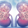
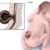
Kidney Cancer
While adenocarcinoma, also called kidney cancer, arises
from small urinary ducts (tubules), a different form called transitional
epithelial cell cancer can develop, which originates in the kidney pool and
ureter canal.
The most common clinical symptoms in kidney cancer are
bleeding in the urine and a palpable mass in the abdomen. There are
usually no symptoms in early stage kidney cancers. And these patients are
usually diagnosed incidentally during ultrasonography or radiological
examinations performed for other reasons.
In the following symptoms, evaluation is required in
terms of kidney tumor;
Bleeding in urine,
A hard mass in the abdomen,
Prolonged and persistent pain in the kidney,
Loss of appetite,
Weight loss
Anemia (anemia).
Abdominal and kidneys should be examined in the diagnosis
and evaluation of kidney cancer. Today, thanks to early diagnosis methods,
kidney tumors can be detected very early and in small scale. For this
reason, “Partial Nephrectomy” in which the kidney is maximally
preserved and only cancerous tissue is removed has become increasingly
important.
Testicular Cancer
Although testicular cancer is a rare type of tumor, it is
the most common cancer in men between the ages of 20-35. There are
situations that are risk factors in the development of testicular cancer.
History of undescended testicles,
Poorly developed testicular structure,
Having a family history of testicular cancer,
Klinefelter syndrome,
The white race.
The most common symptoms of testicular cancer include
hard swelling and a feeling of discomfort in the scrotum. Other symptoms,
such as these mentioned symptoms, may also appear as clinical signs of
testicular cancer. However, it should be kept in mind that some other
diseases may cause similar clinical symptoms. If you have the following
symptoms, you should talk to your doctor without waiting;
Painless swelling in the testicle (egg),
Change in the normal feeling of the testicle,
Dull pain in the lower abdomen or groin,
Sudden accumulation of fluid in the scrotum (scrotum),
Discomfort in the testicles,
Testicular examination and some blood tests are used in
the diagnosis of testicular cancer,
All or some of the tests listed below can be used in the
diagnosis of testicular cancer.
Physical Examination and Story
It is absolutely necessary to have a general examination
of the patient (especially is there a palpable mass in the abdomen?) In
addition, both testicles should be examined carefully, respectively. In
this examination, the general structure of the palpable mass in the testicle
(is it hard, is it irregular in borders? Its approximate size? In what location
in the testicle?) Is evaluated.
Ultrasonography
With this evaluation, the internal structure of the
testis, the features and size of the mass, and the vascular properties of the
tumor if Doppler ultrasonography is performed, are evaluated without bringing
any additional morbidity to the patient.
Serum tumor markers
In testicular cancer, the serum levels of some substances
(markers) secreted by cancer cells are measured. There are 3 tumor markers
in the search for testicular cancer. Alpha-feto protein (AFP), beta-human
chorionic gonadotropin (beta-HCG), lactate dehydrogenase (LDH) These substances
must be checked before radical orchiectomy (removal of the testicle).
Chronic prostatitis
Generally, prostatitis is known as the disease of young
men. However, epidemiological studies show that prostatitis is seen in all
age groups unlike BPH and prostate cancer. Chronic prostatitis begins to
be seen in men as sexual life becomes active. Approximately 5 percent of
men in their 20s to 40-50s experience this ailment. It is the third most
common disease in men over the age of 50. In addition to chronic
prostatitis, burning while urinating and frequent urination, it also causes
pain in the genital area. Chronic prostatitis is not contagious, but it is
a disease that is very difficult to definitively treat and can last for years.
Chronic prostatitis often causes psychological problems
in patients. For this reason, it also affects sexual life
negatively. Pain in the genital areas of men with chronic prostatitis can
also cause impotence anxiety in these people.
What is Chronic Prostatitis?
Prostatitis is an inflammatory disease (inflammation) of
the prostate and sometimes the surrounding tissue and is a prostate problem
frequently encountered by men. Prostatitis is thought to be the cause in 4
out of 10 men who go to a doctor with genital or urinary problems.
The prostate gland, which is found in all healthy men and is necessary for
reproduction, can become inflamed, like any other organ in our
body. Prostate inflammation, or prostatitis, is usually seen in young and
reproductive men. If prostatitis is not treated early, it is very prone to
become chronic.
Prostate inflammation occurs in some prostate types
without bacteria. Prostate inflammation is not one of the sexually
transmitted diseases. It is a condition that can occur at any age after
adolescence.
The diagnosis of chronic (chronic) prostatitis,
which is quite common, may be overlooked because its symptoms are not apparent
and often coexist with other urinary tract inflammations.
Endoscopic treatments in kidney stone disease
Percutaneous nephrolithotripsy (pnl)
As a result of the crystallization of chemical salts in
the urine and the combination of these crystals with each other, kidney stones
or stones occur in the urinary tract. Especially in stones larger than 2
cm in the kidney, there may be problems for the patient in breaking the stone
with the stone crushing device called ESWL or pouring such large stones from
the urinary canals (for example, long and repetitive stone breaking sessions,
insufficient breaking of the stone, incomplete pouring of the broken stones or
pain and obstructions in the kidney, such as stone street, stein strasse, stone
road). As stated in the stone disease diagnosis and treatment guidelines
published by the American Urology Association, the European Association of
Urology and the Turkish Urology Association, the first treatment option for
stones larger than 2 cm in the kidney should be PNL.
Stone treatment with the PNL method is a surgical
treatment that has been applied to many patients all over the world for many
years. It is a reliable method that has replaced open surgery and stone
treatment in almost the majority of patients with kidney
stones. Approximately, surgery takes between 2-4 hours, including
preparation and recovery from general anesthesia. Stone treatment with PNL
method is 1-1.5 cm. It is realized through a hole the size of the
hole. After the intervention, a thin probe (nephrostomy tube) is inserted
into the kidney through the hole where the surgery was performed, and it is
left for 1-2 days for fast postoperative recovery. After PNL, the pain due
to surgery is very short and less, and the duration of hospitalization and the
time to return to daily activities and work is significantly shorter than open
surgery. Usually, the nephrostomy tube is removed on the 1st or 2nd day
after the operation and the patient is removed after observing the cessation of
soaking through the hole for a few hours. A significant cosmetic advantage
is also provided compared to the surgical incision in open stone surgery.
Flexible Ureterorenoscopy (RIRC, Retrograde Intra Renal
Surgery)
The word “flexible” in English means flexible,
flexible. The ureter is the thin channel that carries the urine formed in
the kidney from the kidney pool (pelvis) to the bladder. The ureter is in
two, one in the right and left kidney. The word endoscopy, again from the
English word “Endoscopy”, means looking at an organ in the body
through a telescope. “Flexible Ureterorenoscopy”, which consists
of the combination of these three words, involves entering the ureter and
kidney through the urinary canal through a very thin telescope that can be
folded, without opening any hole in the body, and breaking or removing the
stone or tumor here by laser.
Flexible ureteroscopes that can be folded are usually
8-10 Ch (3.-3.5 mm) diameter thickness. Inside, there is a very thin
working channel used for breaking and removing stones with optical lenses or
digital camera cables. We can easily break the stones in the kidney with
the laser cable system that passes through the thin working channel, and take
the broken large (4-5 mm stone fragments are considered to be large in a study
of this fine) with the basket method. Here, 3 mm and below stone chips are
not collected and can be easily dropped painlessly.